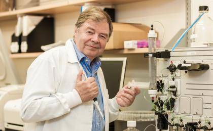
Karl Tryggvason, Tanoto Foundation Professor of Diabetes Research and professor at Duke-NUS Graduate School in Singapore, did not set out to be a world-renowned physician scientist. Instead he started his illustrious career as an architecture student at the University of Oulu in Finland.
Halfway through, he switched to study medicine, which turned out to be his calling. Today his research involves the molecular components, biology and diseases of a special part of the connective tissues: basement membranes (BM). BMs are located closest to most cell types in the body and, as their name suggests, form the base of the biological structure in the body.
Pretty fitting research concentration for an almost architect.
Using his BM expertise, Tryggvason and his team at Karolinska Institutet in Sweden (where he also holds an appointment) recently discovered a method of generating human embryonic stem (hES) cells without destroying any human embryos. Their findings were published this week in the journal Nature Communications.
The two main hurdles for hES cell applications have been ethical and technical. The ethical concern is that it is generally believed that one cannot generate hES cell lines without destroying human life, which is why it is so difficult to get approval for the generation of hES cell lines in the United States. Using a standard IVF procedure, Tryggvason and his team demonstrated that it is indeed possible to clonally derive new hES lines without destroying an embryo.
The technical issue was that until now there hasn't been a chemically defined, xeno-free method for deriving and expanding hES cells. The researchers solved this by using the human xeno-free laminin substrate (LN-521) and adhesion molecule found to associated with pluripotent stem cells in the early human embryo. Tryggvason's team produced this protein in the laboratory and isolated it for culturing the hES cells.
"It is my hope that the results of this study will help soften up negative attitudes toward the establishment and use of hES cells for the development of cell therapy in regenerative medicine," said Tryggvason.
A central part of the discoveries have to do with use of laminins -- a family of base proteins. Early in his career, the first laminin protein was isolated at the National Institute of Health, where Tryggvason was a postdoc. Realizing later that there are at least 16 in the body, he questioned why there were so many. He presumed they were important or phenotypically specific to the specific cell types and organs they supported. He was right.
Tryggvason has a large body of data showing that specific laminins support cellular phenotypes. Using this knowledge he created in vitro environments that mimic those in vivo and it helped to develop better protocols for cell differentiation and phenotype stability, especially in the case of the hES cell generation.
"Our findings enable the robust expansion of hES cells and allow us to envision the establishment of a hES cell bank that could represent most tissue antigen classes is possible. A stem cell bank could mean that in the future, these cells that are representative of different tissue antigens, etc., could be stored for use," Tryggvason said. "Imagine the limitless possibilities of their use in regenerative medicine."
CITATION: Clonal culturing of human embryonic stem cells on laminin-521/E-cadherin matrix in defined and xeno-free environment, Rodin S. et al. Nature Communications. Online Jan. 27, 2014. doi:10.1038/ncomms4195